One part of the skeleton reconstruction I found challenging was the
pelvis. The drawings of Madsen (1976) are complete but not easy to
follow. Each sacral vertebrae and rib is shown separately from front,
back and side. However, how they all articulate is not shown and I struggled to
understand how all the bones joined up. So I fitted them together virtually on the computer screen.
Each of Madsen's lateral drawings was scanned, rotated, cleaned, and scaled into separate layers in PhotoShop. I could then experiment with how to fit them together and also hide/show different bones. The sacral ribs are hard to see, so these were recolored for contrast
For larger elements (ilium, pubis and ischium), this method allowed me to get an overall picture of the pelvic assembly, a view that was not offered by Madsen.
Madsen's drawings are composite drawings made from different individuals. This means the complex suture and scar joints of the different bones are representative, but do not correspond exactly bone-to-bone. This was not obvious to me at the start, and you will notice those inconsistencies if you look closely. It made exact placement of sacral ribs especially difficult and they may not be exactly correct as shown here.
Update
Fernando Novas
visited recently and I discussed the
Allosaurus pelvis with him. He kindly sent me
scanned images from the original Gilmore 1920 manuscript in his collection [Gilmore, Charles W. (1920). "Osteology of the carnivorous dinosauria in the United States National Museum, with special reference to the genera Antrodemus (Allosaurus) and Ceratosaurus". Bulletin of the United States National Museum 110: 1–159]. I cleaned the
scans and appended the separate figure legends to each. See them below.
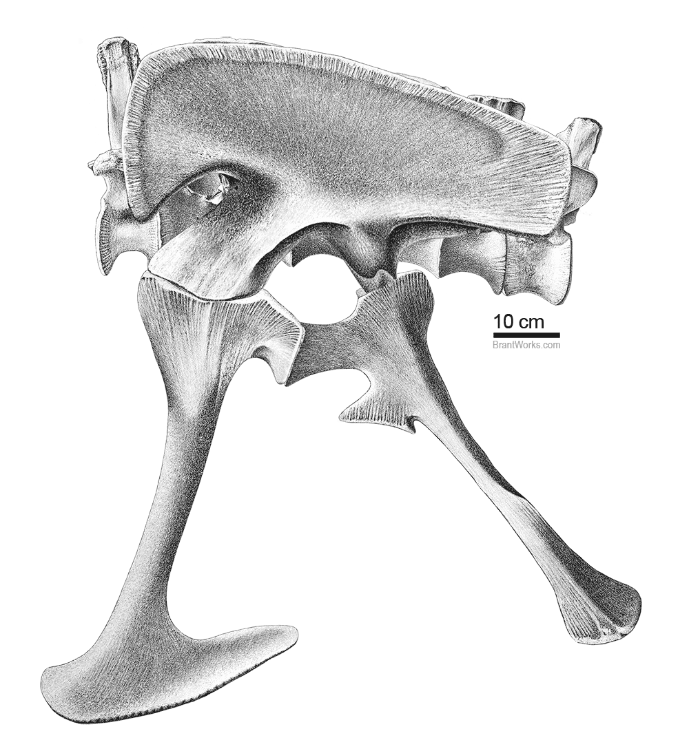
Fig 1
Full assembly including the last dorsal vertebrae (D14) and the first Caudal (C1)
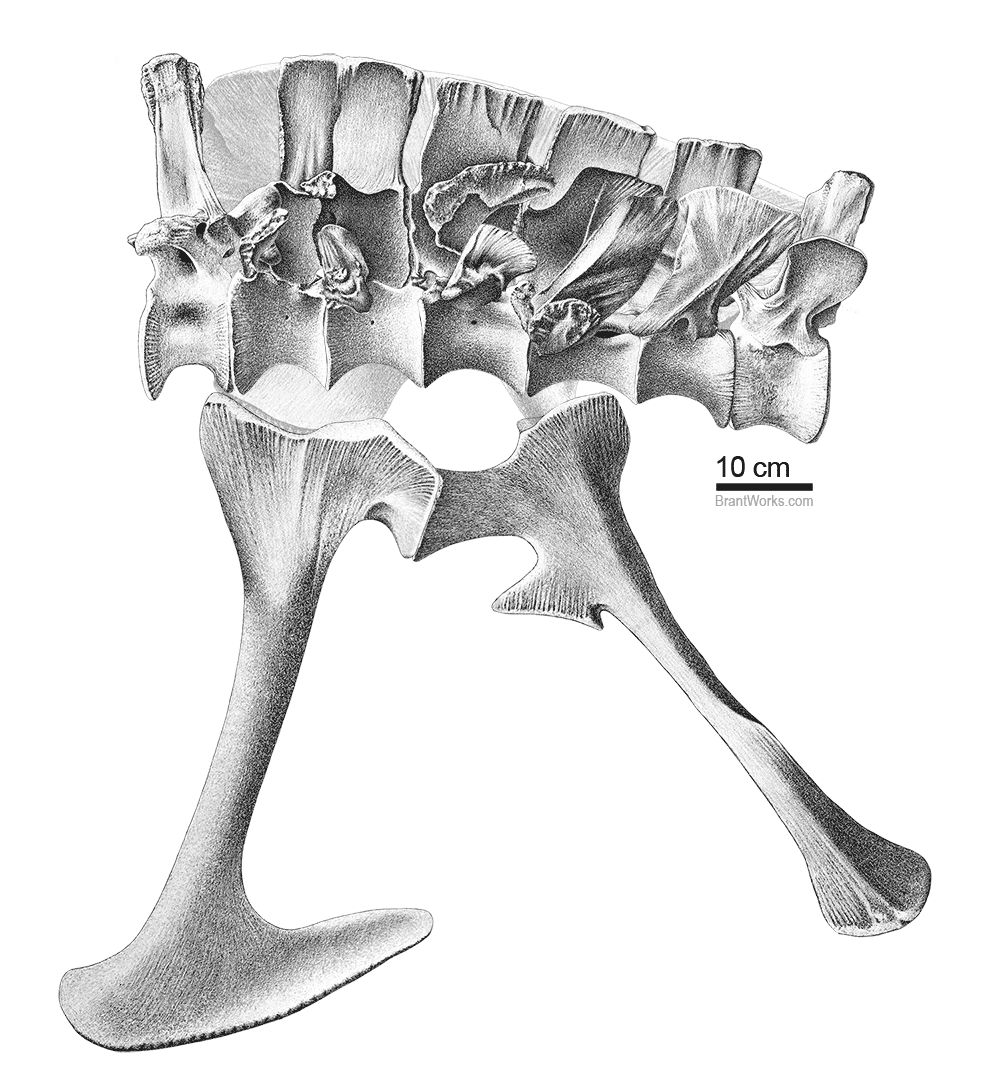
Fig 2
Full assembly including the last dorsal vertebrae (D14) and the first Caudal (C1). The ilium has been removed to show articulation of all the sacral vertebrae and sacral ribs.

Fig 3
Major bones in articulation; the pubis, ischium and ilium. The back of the ilium is shown so scarring from the points of contact (attachment) with the sacral vertebrae and ribs can be seen.
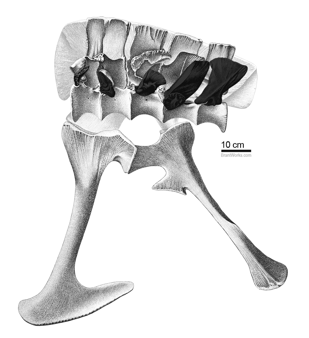
Fig 4
Full
pelvic assembly. The proximal ilium has been removed to show articulation of all the
sacral vertebrae and ribs. Sacral ribs are darkened for contrast.
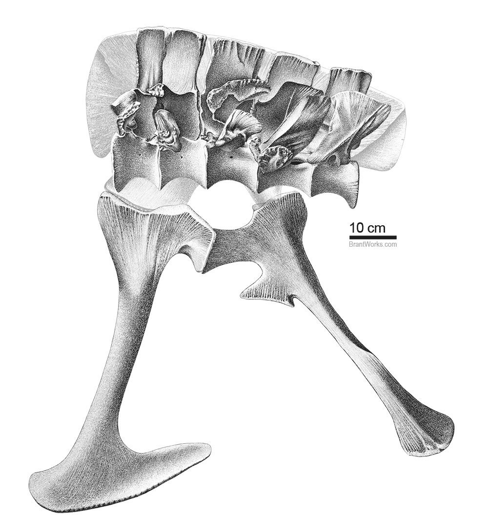
Fig 5
Full
pelvic assembly. The proximal ilium has been removed to show articulation of all the
sacral vertebrae and ribs.
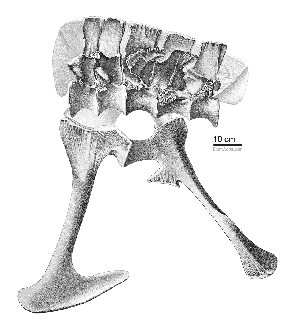
Fig 6
Full
pelvic assembly. The proximal ilium and sacral ribs have been removed to show articulation of the
sacral vertebrae.
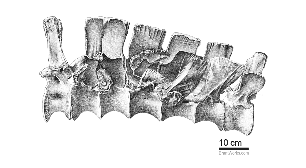
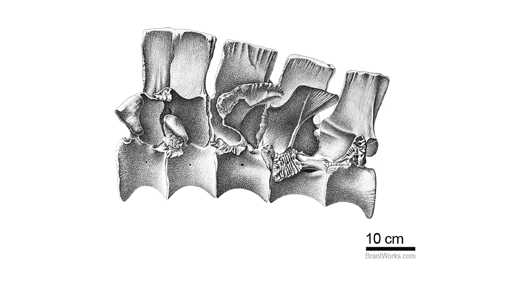
Fig 7
Articulation of sacral vertebrae and sacral ribs including the last dorsal vertebrae (D14) and the
first Caudal (C1). The large gap between the neural spines of the last dorsal and first sacral was surprising. It makes an obvious breakpoint between the dorsal and the sacral region of the spinal column.

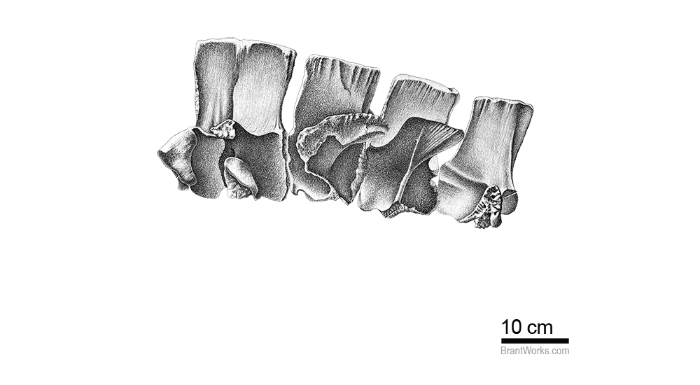
Fig 8
Articulation of sacral vertebrae and sacral ribs. Sacral ribs are darkened for contrast.
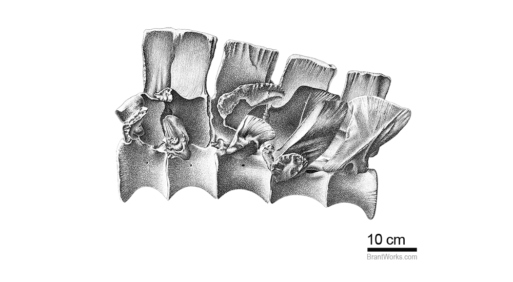
Fig 9
Articulation of the sacral vertebrae and sacral ribs.
A
B
A
B

Fig 10
Articulation of sacral vertebrae. The sacral ribs have been removed to show articulation of each vertebrae.
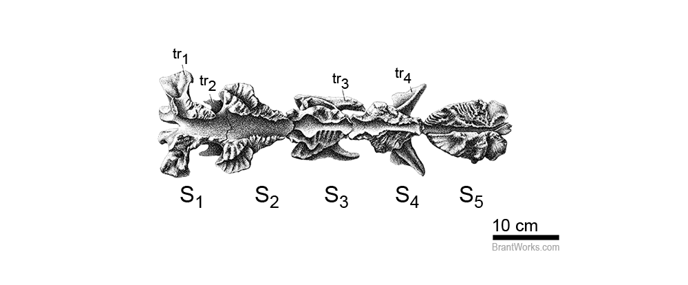
Fig 11
Articulation of sacral neural spines in A; lateral view and B; ventral view. Ventral articulation is from Madsen (1976). Abbreviations: S1-5 = sacral 1-5; tr1-4 = transverse process 1-4.
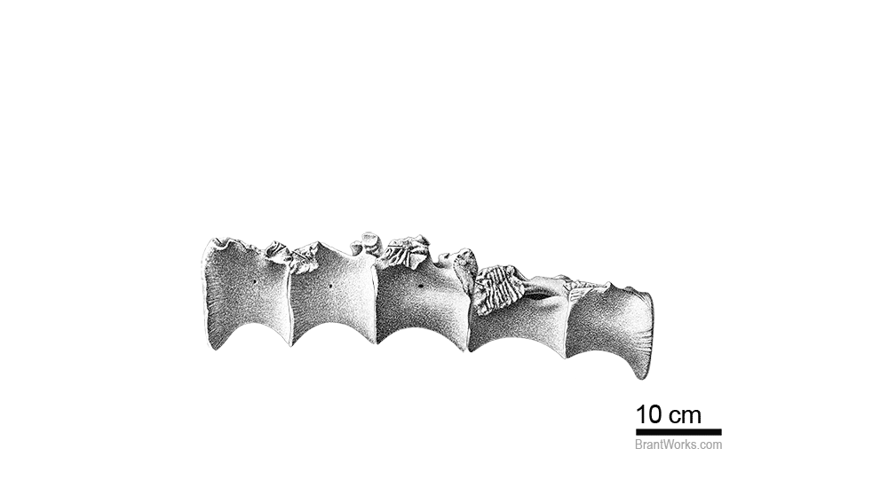
Fig 12
Articulation of sacral centra in A; lateral view and B; dorsal view. Dorsal articulation is from Madsen (1976). Abbreviations: S1-5 = sacral 1-5; tr1-4 = transverse process 1-4; nc = neural canal.
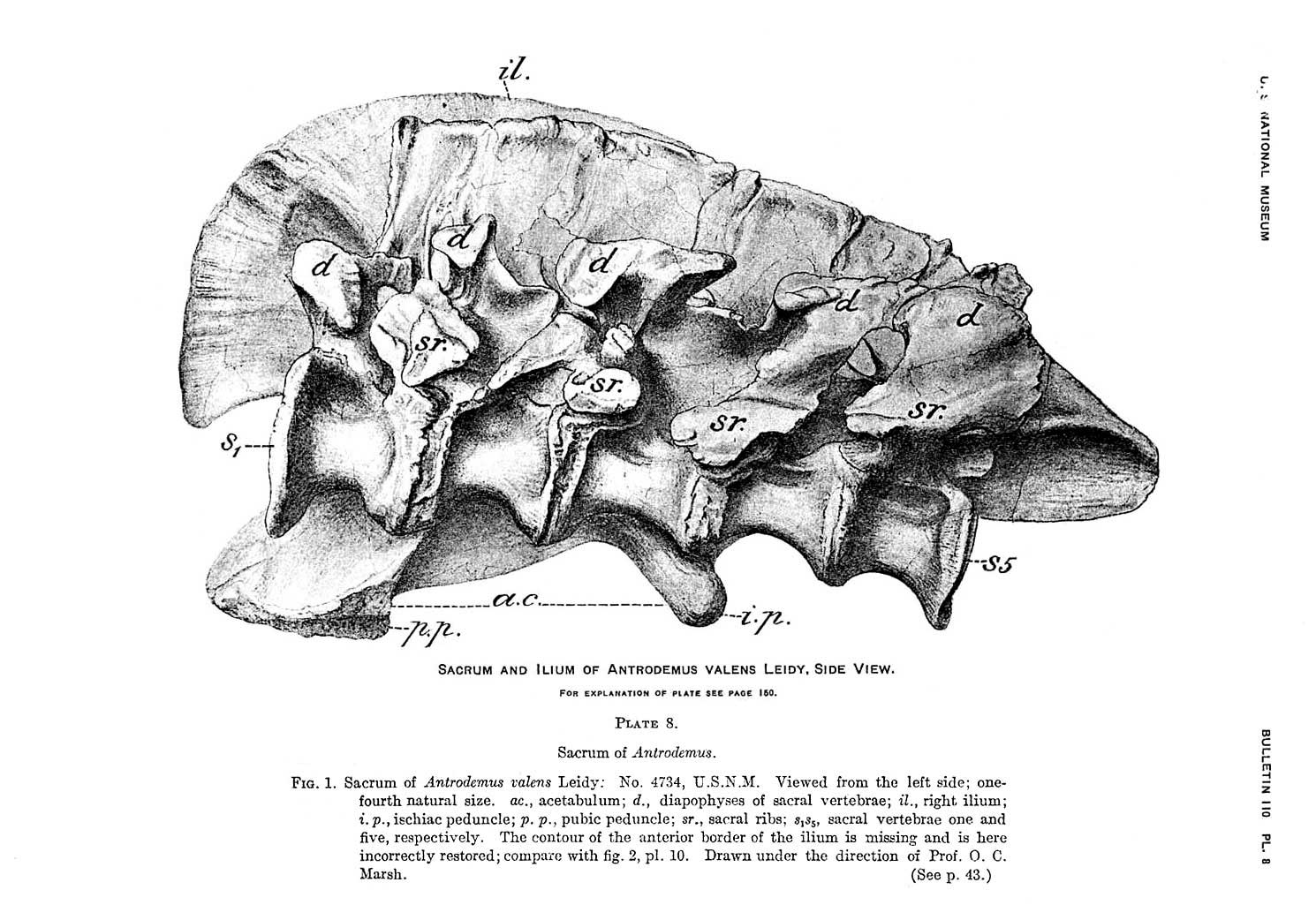
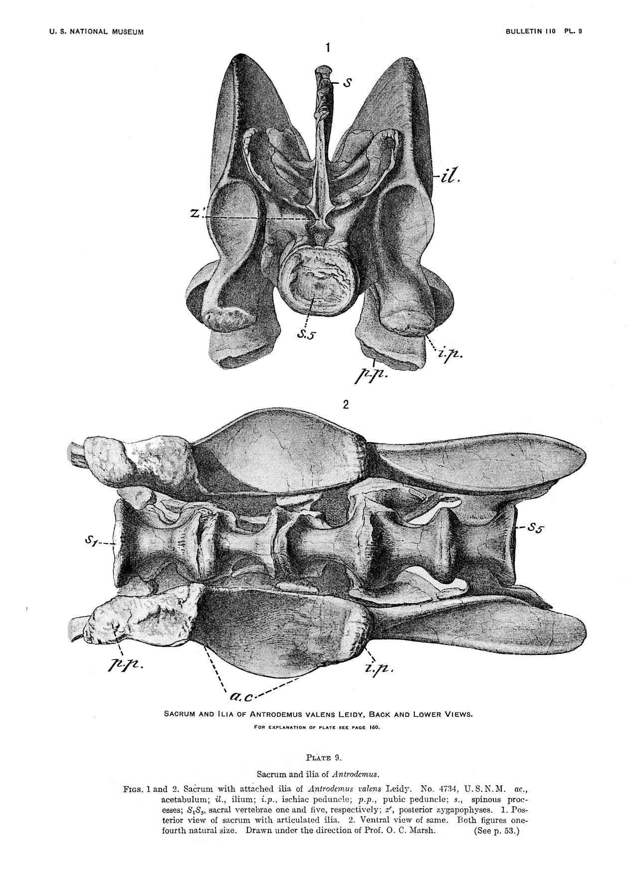
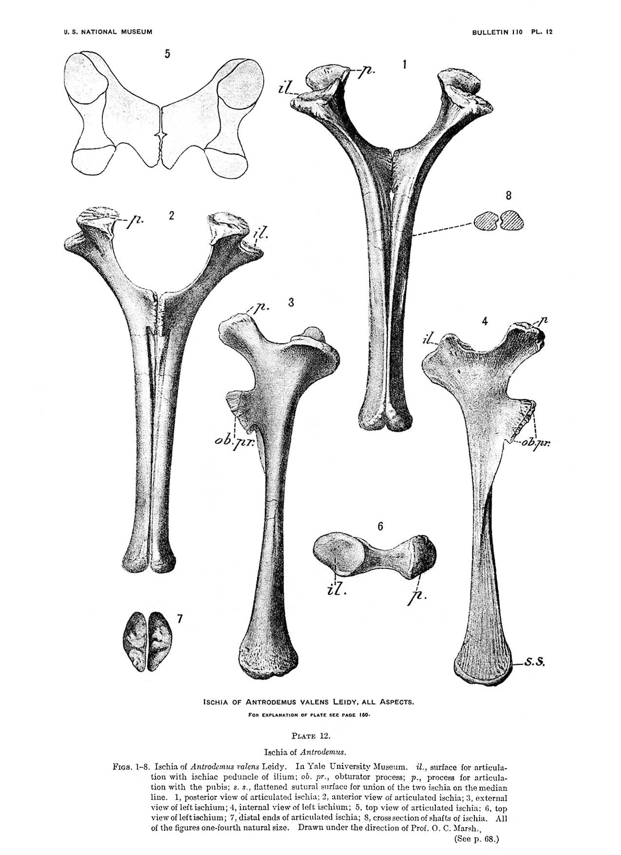
The following images are from the manuscript of Gilmore, Charles W. (1920). "Osteology of the carnivorous dinosauria in the United States National Museum, with special reference to the genera Antrodemus (Allosaurus) and Ceratosaurus". Bulletin of the United States National Museum 110: 1–159. The drawings in Gilmore's publications are always superb! Click each image to view full size.