Images I found most useful when researching the skeleton:
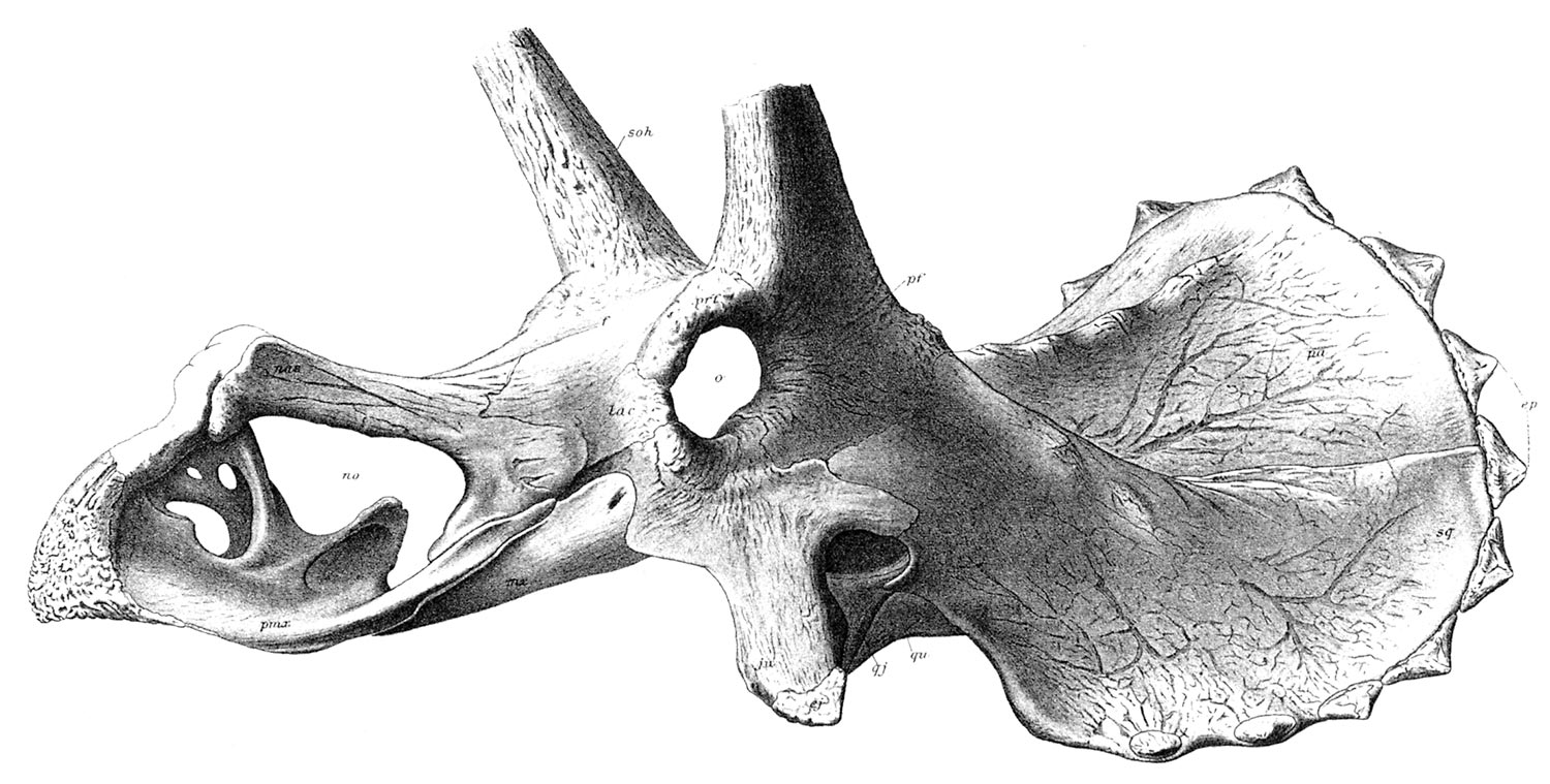
Triceratops serrtus skull
From Hactcher et al 1907, The Ceratopsia, United States Geological Survey Monograh 49: 1-300.
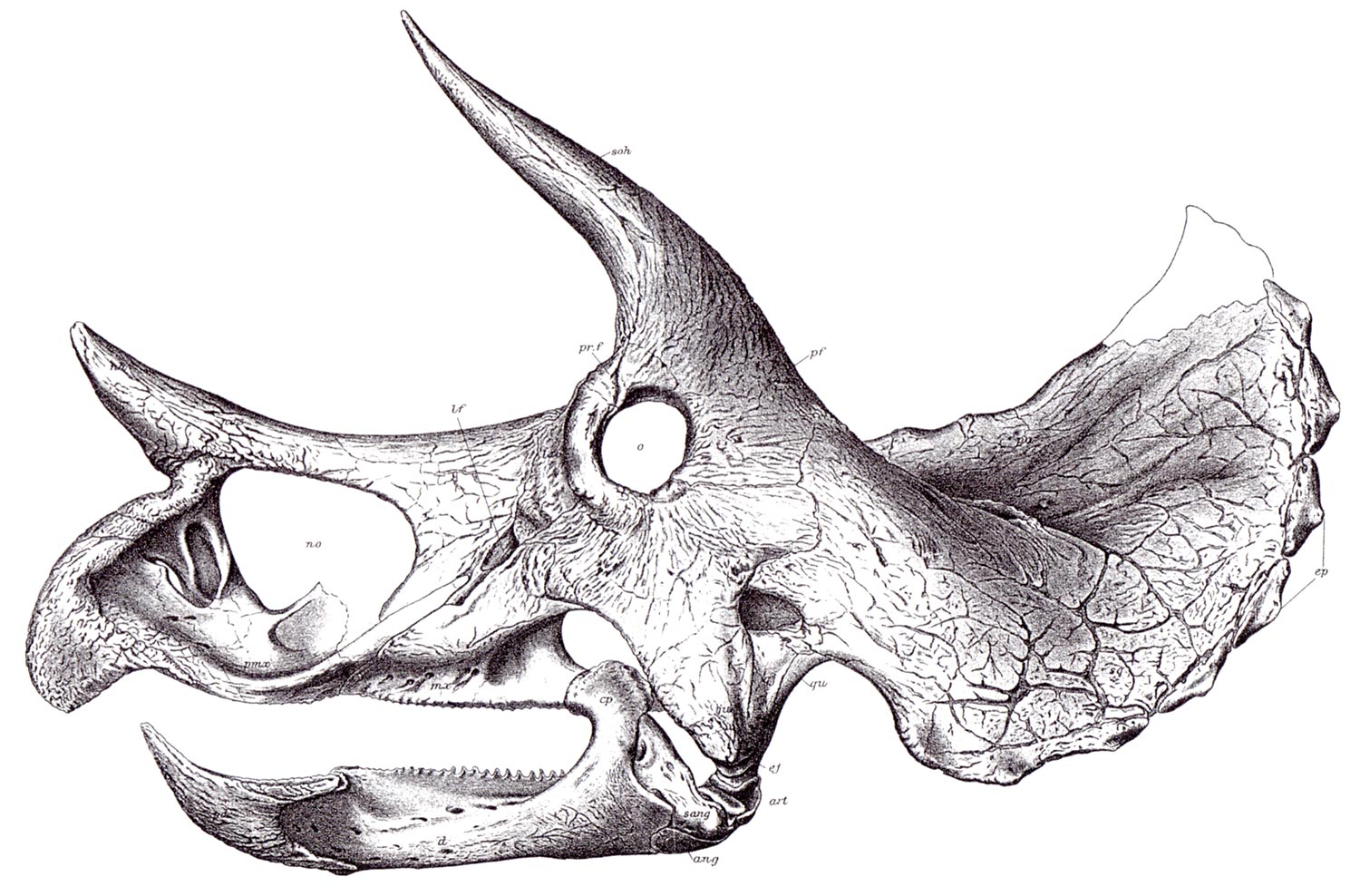
Triceratops prorsus skull
From Hactcher et al 1907, The Ceratopsia, United States Geological Survey Monograh 49: 1-300.

Occipital condyle (Top) and side view of same showing braincase
Abbreviations; foramen magnum (fm), basioccipital (bo), fossa ovale (fo), opening for
internal carotid artery (lca). Roman numerals indicate entry points for cranial nerves to
the brain. From Dodson P. 1996. The horned Dinosaurs, Princeton University Press.
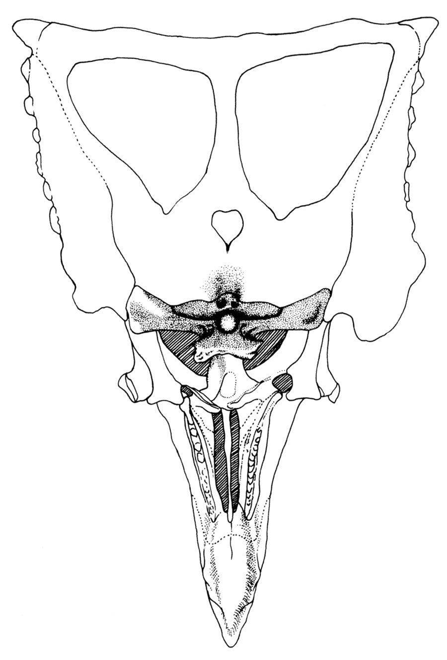
Underside of Chasmosaurus belli skull showing occipital condyle
From Dodson P. 1996. The horned Dinosaurs, Princeton University Press.
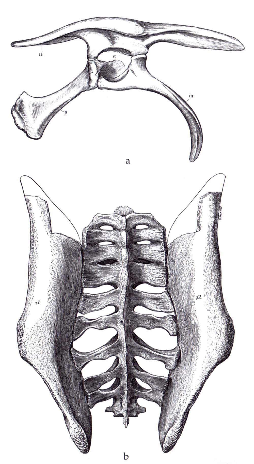
Triceratops pelvis in (a) lateral and (b) dorsal views.
Abbreviations; (il) ilium, (p) pubis, (is) ischium, (a) acetabulum.
From Marsh O. C 1891, Restoration of Triceratops. American Journal of Science, 41:339-342
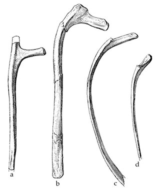
Triceratops ribs
Representative ribs from Brachyceratops (a) second dorsal, (b) middle dorsal,
(c) another middle dorsal, (d) final dorsal. From Gilmore C.W. 1917 Brachyceratops,
a ceratopsian dinosaur from the Two Medicine Formation of Montana with notes
on associated reptiles. U.S. Geological Survey Professional Paper 103: 1-45.
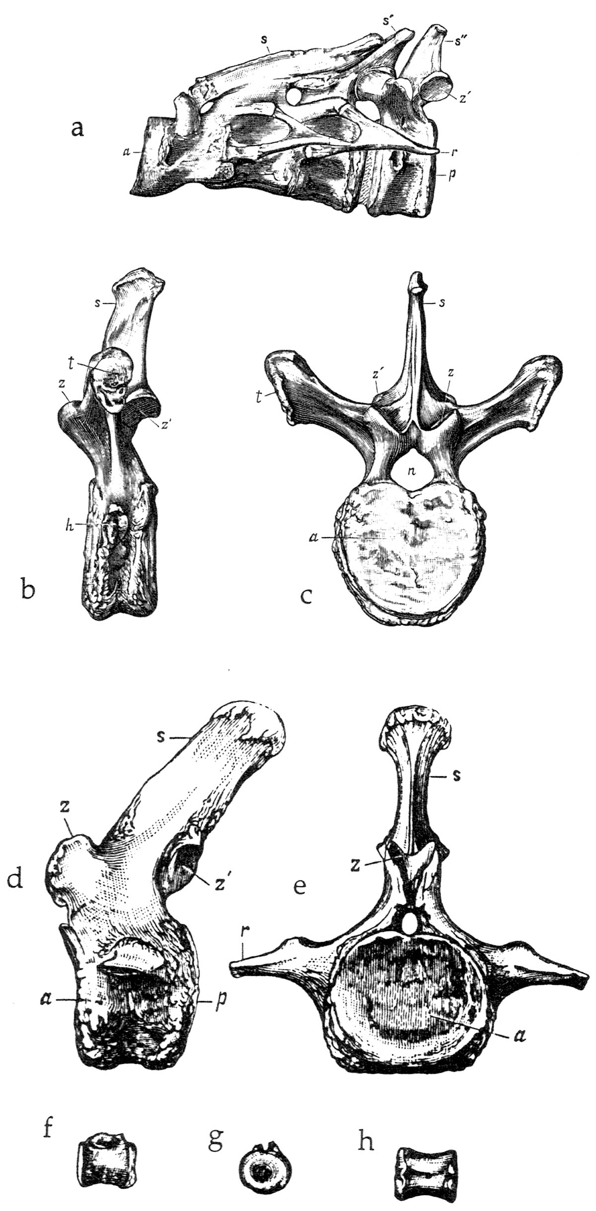
Triceratops vertebrae
(a) Syncervical, or fused cervical (neck) vertebrae (i.e. atlas/axis complex; vertebrae 1-4 inclusive),
(b) & (c) second dorsal vertebrae, (d) & (e) first caudal (tail) vertebrae (f), (g), & (h) distal caudal vertebrae.
From Marsh O. C 1891, The gigantic Ceratopsidae, or horned dinosaurs, of North America. American Journal of Science, 41:167-178